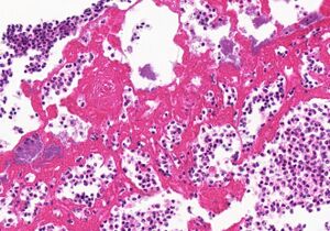
Description: The slide is from a cross-section of the heart. Five layers can be distinguished. From the bottom layer to the top layer:
The bottom layer is the myocardium. It’s normal.
The second layer is the visceral pericardium. It’s also normal.
The middle layer contains fibrosis
The second-from-the-top layer is pale, has some neutrophils and contains some loose connective tissue
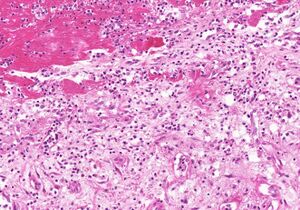
Close-up of layer 5. The red hyaline is fibrin, and the leukocytes are neutrophils.Close-up of layer 4. You can see loose connective tissue and fibrin. In the top left you can see a part of the fifth layer.The top layer is very eosinophilic with a glass-like (hyaline) structure. It contains many neutrophils.
Diagnosis: Fibrinous pericarditis = cor villosum
Theory: The fibrinous exudate caused by the fibrinous acute inflammation is mostly degraded by fibrinolysis and macrophages, a process called resolution. However, the fibrin-rich exudate is not completely removed and is instead replace by fibroblasts and blood vessels invading the exudate to form fibrosis. This is the process called organization.
The fifth layer (layer 5) shows the active phase of the pericarditis. It’s comprised of only neutrophils (due to the inflammation) and fibrin. Fibrin, being a protein, is very eosinophilic under the microscope. The fourth layer shows the early phase of organization after the pericarditis. This layer is where organization happens. It is paler than the fifth layer because this layer doesn’t contain just fibrin, but loose connective tissue as well, made by invading fibroblasts. The third layer shows the late phase of organization. In this layer, fibrosis has already started. The second and first layers are normal. The fibrous scar tissue forming in the pericardium can restrict the function of the myocardium.