File:Coeliac disease - overview.png

Size of this preview: 734 × 600 pixels. Other resolution: 1,796 × 1,468 pixels.
Original file (1,796 × 1,468 pixels, file size: 2.55 MB, MIME type: image/png)
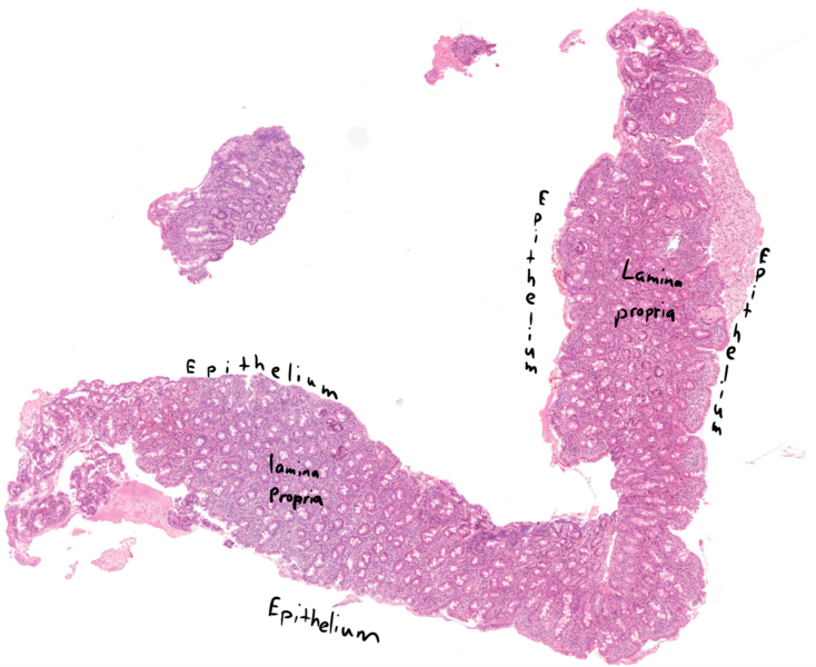
Overview. In most of the epithelium there is complete villous atrophh, as no villi are visible – the mucosa is almost completely flat. On the epithelium on the right side you can see some very atrophic villi (see the comments). Even with low magnification can we see that the number of crypts is high compared to normal.
File history
Click on a date/time to view the file as it appeared at that time.
| Date/Time | Thumbnail | Dimensions | User | Comment | |
|---|---|---|---|---|---|
| current | 10:24, 16 June 2022 | | 1,796 × 1,468 (2.55 MB) | Nikolas (talk | contribs) |
You cannot overwrite this file.
File usage
The following page uses this file: