File:The light-microscopical changes in necrosis..png

Size of this preview: 774 × 600 pixels. Other resolution: 1,474 × 1,142 pixels.
Original file (1,474 × 1,142 pixels, file size: 3.24 MB, MIME type: image/png)
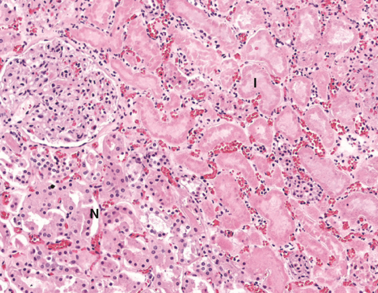
The light-microscopical changes in necrosis. Taken fron a kidney infarction. «I» shows the infarcted area, while «N» shows the normal tissue. Notice that the infarcted tissue is more eosinophilic, has less structure and has fewer nuclei.
File history
Click on a date/time to view the file as it appeared at that time.
| Date/Time | Thumbnail | Dimensions | User | Comment | |
|---|---|---|---|---|---|
| current | 10:10, 6 May 2022 | | 1,474 × 1,142 (3.24 MB) | Nikolas (talk | contribs) |
You cannot overwrite this file.
File usage
The following page uses this file: