(Created page with "'''Staining''': HE '''Organ''': Parotid gland '''Description''': The bottom left half of the slide shows the normal fat rich, serous parotid gland. The upper right half is the tumor. It consists of glandular structures and epithelial-like cells. There are also some areas inside the tumor with cartilage-like tissue. '''Diagnosis''': Pleomorphic adenoma '''Causes''': * Chromosomal rearrangements involving PLAG1 gene (not important) '''Theory''': This benign neopla...")
* [[File:Pleiomorphic adenoma - high magnification.png|thumb|Basically every cell you can see around the cartilage is a ductal epithelial cell. You don’t have to identify a myoepithelial cell.|346x346px]]Chromosomal rearrangements involving PLAG1 gene (not important)
'''Theory''':
'''Theory''':
Line 24:
Line 24:
Pleomorphic adenoma usually grows small “feet”, called pseudopods, taking on a shape similar to a starfish with short legs. One pseudopod can be seen on the slide.
Pleomorphic adenoma usually grows small “feet”, called pseudopods, taking on a shape similar to a starfish with short legs. One pseudopod can be seen on the slide.
[[File:Pleiomorphic adenoma - overview.png|center|thumb|Overview of the slide]]
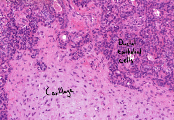
[[File:Pleiomorphic adenoma - high magnification.png|center|thumb|Basically every cell you can see around the cartilage is a ductal epithelial cell. You don’t have to identify a myoepithelial cell.]]
[[Category:Pathology 2 - Histopathology slides]]
[[Category:Pathology 2 - Histopathology slides]]
Latest revision as of 13:10, 7 July 2024
Overview of the slide
Staining: HE
Organ: Parotid gland
Description:
The bottom left half of the slide shows the normal fat rich, serous parotid gland.
The upper right half is the tumor. It consists of glandular structures and epithelial-like cells. There are also some areas inside the tumor with cartilage-like tissue.
Diagnosis: Pleomorphic adenoma
Causes:
Basically every cell you can see around the cartilage is a ductal epithelial cell. You don’t have to identify a myoepithelial cell.Chromosomal rearrangements involving PLAG1 gene (not important)
Theory:
This benign neoplasm gets its name from the pleomorphism it has. It consists of a mixture of ductal epithelial cells, myoepithelial cells and mesenchymal tissue like cartilage, loose connective tissue or even bone. We can therefore say that this tumor has both epithelial and mesenchymal differentiation.
This is the most common benign salivary gland tumor, accounting for around 80% of them. It’s most commonly found in the parotid but can be found in the submandibular or minor salivary glands as well. It usually presents as a palpable mass.
Treatment involves surgical removal, however complete removal is difficult because of the anatomical relationship with the facial nerve. This tumor has a high recurrence rate if not removed completely. It also has a high risk of turning malignant, turning into carcinoma ex pleomorphic adenoma, also called CEPA.
Pleomorphic adenoma usually grows small “feet”, called pseudopods, taking on a shape similar to a starfish with short legs. One pseudopod can be seen on the slide.