(Created page with "'''Staining''': HE '''Organ''': Brain '''Description''': You can clearly see an area that is different from the rest. This area has no nuclei, is pale and has loose parenchyme. There are hypereosinophilic neurons with karyopyknosis (shrinkage of the nuclei). '''Diagnosis''': Encephalomalacia alba '''Theory''': There are three types of encephalomalacia. Alba (white), flava (yellow) and rubra (red). Alba is “fresher”, more recent than the flava type, and will bec...")
[[File:Encephalomalacia alba overview.jpg|thumb|Guess which area is important here]]'''Staining''': HE
'''Organ''': Brain
'''Organ''': Brain
Line 17:
Line 17:
The alba and flava are caused by a blockage of cerebral artery by:
The alba and flava are caused by a blockage of cerebral artery by:
* Thrombosis
* [[File:Encephalomalacia alba overview infarct.jpg|thumb|The hypereosinophilic cells are the red neurons.]]Thrombosis
* Embolization
* Embolization
* Vasculitis
* Vasculitis
Line 26:
Line 26:
* Cerebral tumor
* Cerebral tumor
* Cerebral abscess
* Cerebral abscess
[[File:Encephalomalacia alba overview.jpg|center|thumb|Guess which area is important here]]
[[File:Encephalomalacia alba overview infarct.jpg|center|thumb|The hypereosinophilic cells are the red neurons.]]
[[Category:Pathology 1 - Histopathology slides]]
[[Category:Pathology 1 - Histopathology slides]]
Latest revision as of 11:12, 5 July 2024
Guess which area is important here
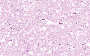
Staining: HE
Organ: Brain
Description:
You can clearly see an area that is different from the rest. This area has no nuclei, is pale and has loose parenchyme. There are hypereosinophilic neurons with karyopyknosis (shrinkage of the nuclei).
Diagnosis: Encephalomalacia alba
Theory:
There are three types of encephalomalacia. Alba (white), flava (yellow) and rubra (red). Alba is “fresher”, more recent than the flava type, and will become the flava type over time. The name of the rubra comes from the bleeding that accompanies it. The first two are examples of anemic “infarction” while the rubra is a type of haemorrhagic “infarction”.
The hypereosinophilic neurons are called red neurons. Neurons are very sensitive to hypoxia, so they are the first to be damaged during hypoxia.
The alba and flava are caused by a blockage of cerebral artery by:
The hypereosinophilic cells are the red neurons.Thrombosis
Embolization
Vasculitis
The rubra type is caused by venous obstruction by: