8. Steatosis hepatis
Staining: HE and oil-red

Organ: Liver
Description:
HE: We can see the portal triad, meaning that we are looking at a liver. There is panlobular fat accumulation (fat is present in the whole lobule) in the whole slide.
Oil-red: With this staining the fat is reddish-orangeish.
Diagnosis: Hepatic steatosis
Causes:
- Alcohol consumption
- Metabolic syndrome (DM2, obesity)
- Medications
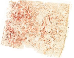
Overview of oil red

Theory:
When fixating the normal slides, we use formalin. Formalin dissolves fat, which is why fat isn’t directly seen in the HE slides. In the oil-red slide there was no formalin but instead the slide was frozen. This preserves the fat.
Oil-red can be used to determine the amount of fat and therefore the severity of the steatosis.
In this slide there was panlobular fat, but in some cases there can be pericentral fat, where fat can only be found around the central veins.
The enzyme ADH that metabolizes ethanol depletes NAD+ and increases NADH. NADH stimulates fatty acid synthesis. NAD+ is needed for β-oxidation of the fat. This causes fat to accumulate in the hepatocytes.


