File:Muscle fibre microanatomy.png
Size of this preview: 748 × 600 pixels. Other resolution: 801 × 642 pixels.
Original file (801 × 642 pixels, file size: 334 KB, MIME type: image/png)
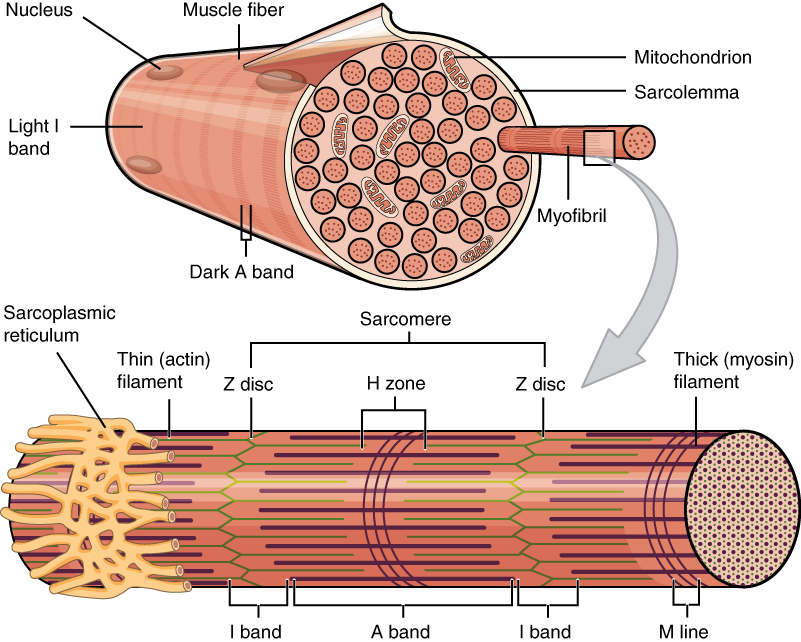
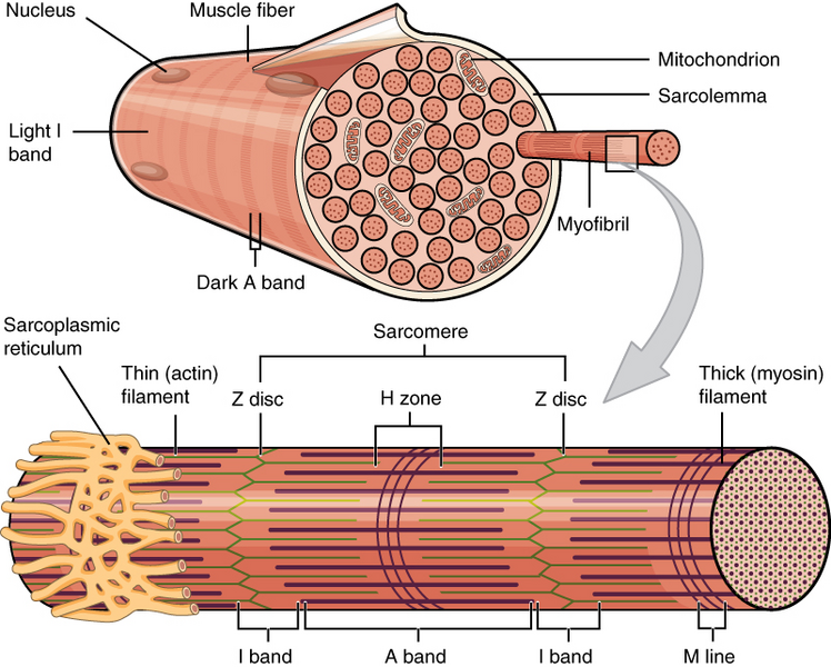
This image shows one muscle fibre in cross-section. In this section it’s easy to see the individual myofibrils which make up the muscle fibre. Thin filament in green and thick filament in reddish-brown something. From https://en.wikipedia.org/wiki/Muscle_contraction
File history
Click on a date/time to view the file as it appeared at that time.
| Date/Time | Thumbnail | Dimensions | User | Comment | |
|---|---|---|---|---|---|
| current | 22:19, 30 November 2022 | | 801 × 642 (334 KB) | Nikolas (talk | contribs) |
You cannot overwrite this file.
File usage
The following page uses this file: